


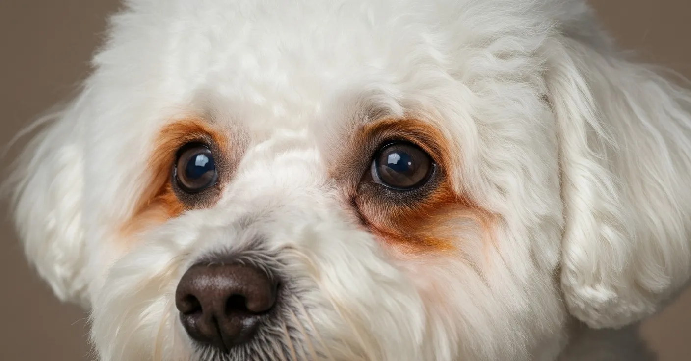
A woman in my neighborhood adopted a Bichon Frise last spring. The dog came from a rescue — surrendered by an elderly owner who couldn’t care for him anymore — and the first thing she noticed at pickup was the heavy rust staining beneath both eyes. She assumed it was neglect. Weeks of missed grooming, maybe a low-quality diet, something the previous owner hadn’t kept up with. She came home determined to fix it.
By week three, after daily face wipes, a switch to premium food, and filtered water in a stainless steel bowl, the staining was already coming back. Just as dark. Just as symmetrical. She called the rescue thinking something was wrong with the dog. Nothing was wrong with the dog. He was a Bichon Frise doing exactly what Bichon Frises do.
That’s the thing owners of these breeds eventually learn: the staining isn’t a failure of care. It’s anatomy. It’s coat chemistry. It’s the predictable intersection of skull shape, eye prominence, and the particular way white fur behaves when it’s exposed to oxidized porphyrins. Understanding which breeds are most affected — and why — sets realistic expectations and points toward the management approaches that actually help.
Why white coats make staining visible
Let’s start here, because it’s a misconception worth clearing up early. White fur doesn’t cause tear staining. It reveals it.
Dark-coated dogs produce porphyrin in their tears at the same rate. They experience the same overflow, the same oxidation, the same microbial activity at the eye margin. The difference is purely visual contrast. Rust-brown on black fur is invisible. Rust-brown on white fur looks like a paint spill. If you could somehow strip the pigment from a black Labrador’s coat, you’d find staining at the inner corners of both eyes.
This distinction matters practically because it explains why interventions aimed purely at whitening the coat don’t solve the problem — they just bleach what’s there temporarily. And it means that when we talk about white-coated breeds being “stain-prone,” we’re really talking about breeds where staining is visible, not where it’s more severe in an absolute biochemical sense. Though as we’ll see, several white-coated breeds also have genuine anatomical risk factors that do increase tear overflow — so visibility and actual volume often compound each other.
The two anatomical risk factors
Across all the breeds discussed in this article, two structural features drive most of the increased staining burden.
Brachycephalic skull shape. Selective breeding for shorter, flatter faces compresses the nasolacrimal duct — the narrow channel that normally drains tears from the inner corner of the eye down through the facial bones and out near the nostril. In a skull built for that drainage path, it works efficiently. In a compressed skull, the duct runs a distorted course: kinked, narrowed, or angled in ways it wasn’t designed for. Tears that should drain internally instead overflow onto the face. This is structural epiphora — excessive tear spillage caused by anatomy, not disease (Gelatt et al., 2013).
Prominent, forward-set eyes. Several breeds — some brachycephalic, some not — have slightly exophthalmic (protruding) eyes. Prominent eyes expose more ocular surface to air, which increases evaporative tear loss and stimulates reflex tearing as compensation. They also allow less eyelid contact with the ocular surface, which can impair the normal spread and drainage of the tear film. More tears on the surface means more tears spilling onto the fur (Maggs et al., 2013).
Many white-coated breeds have one of these features. Several have both. And those that have neither still present staining as a visibility issue simply because of their coat color.
Breed-by-breed breakdown
Maltese
The Maltese is probably the first breed most people picture when they hear “tear stains,” and for good reason. The flowing white coat, the dark eyes, the delicate facial structure — it’s a combination almost designed to display porphyrin staining at maximum contrast.
Interestingly, the Maltese isn’t the most structurally compromised breed on this list. Their nasolacrimal anatomy is relatively normal compared to the hard brachycephalics. What distinguishes them is the coat itself: long, fine, and pale, it lies close to the skin beneath the eyes and holds moisture against the face for extended periods. Oxidation happens right there, in contact with the skin, in a warm environment. The staining tends to run deep and dense on Maltese for this reason.
Many Maltese owners also notice staining at the corners of the mouth — salivary porphyrin deposits from normal self-grooming. Managing this means keeping the muzzle area dry and considering the porphyrin load systemically, not just at the eyes (Petersen-Jones & Crispin, 2002).
Bichon Frise
Bichons present a slightly different challenge. The coat is curly and dense rather than long and silky, which means moisture gets trapped in the curl structure rather than lying flat against the skin. The result is a humid microenvironment at the eye margin that’s highly hospitable to Malassezia yeast — and yeast metabolites stack on top of porphyrin discoloration to produce staining that’s darker and more tenacious than porphyrin alone would generate.
Bichons also carry moderate brachycephalic anatomy. Not as extreme as a Shih Tzu or a Pug, but enough to impair nasolacrimal drainage in a meaningful number of individuals. The combination of drainage impairment and moisture-trapping curl makes them one of the more consistently affected breeds in practice.
Shih Tzu
If you want to understand brachycephalic tear staining at its most consistent, look at the Shih Tzu. The breed standard calls for a broad, round skull and a very short muzzle — the nasolacrimal duct takes a severe detour through that compressed geometry, and significant drainage impairment is nearly universal in the breed. Studies of nasolacrimal anatomy in dogs consistently use brachycephalic breeds — with Shih Tzus frequently represented — as examples of structural epiphora (Bedford, 1999).
Shih Tzus also have large, prominent eyes that are relatively exposed, increasing evaporative tearing and making drainage even less adequate relative to production. Owners of Shih Tzus should understand from the start that staining management is going to be a consistent, ongoing routine rather than a problem to be solved once and set aside. That’s not pessimism; it’s anatomy.
Toy and Miniature Poodle
Poodles bring a different risk profile to the table. They’re not brachycephalic — the skull is long and the nasolacrimal anatomy is typically intact. What Poodles carry is a genetic predisposition to epiphora that appears to run through certain lines, particularly in the Toy and Miniature size variants. The mechanism isn’t fully characterized, but elevated baseline tear production is a consistent finding in affected individuals.
The coat compounds the issue. Light-colored curly Poodle coat — especially the apricot, cream, and white varieties — traps moisture and resists drying in the same way Bichon coat does. Rust-brown staining shows clearly against the pale background. The good news is that Poodles without the structural epiphora of brachycephalic breeds often respond well to dietary adjustments and supplementation targeting porphyrin load, since their problem is more about volume and chemistry than anatomy (Maggs et al., 2013).
Cocker Spaniel
The American Cocker Spaniel deserves a separate mention because it brings a risk factor the others don’t: eyelid conformation issues. Entropion — where the eyelid rolls inward so that the lashes contact the corneal surface — is more common in Cockers than in most other breeds on this list. Entropion causes chronic corneal irritation, which drives reflex tearing as a protective response. That’s pathological epiphora: tear overflow caused by active irritation, not just drainage impairment.
Owners of Cockers with heavy staining should specifically have a vet evaluate eyelid conformation. Correcting entropion surgically can significantly reduce tear overflow and, by extension, staining. Supplementation and grooming help manage the staining, but they don’t address the underlying lid abnormality if one is present (Stades et al., 2007).
West Highland White Terrier
Westies aren’t brachycephalic, and they don’t typically have the extreme eye prominence of some breeds on this list. What they have is a combination of deep-set eyes — which can impair tear drainage by changing the geometry of the lower lid and punctum — and a very white, dense double coat that shows every trace of discoloration.
Westies also tend to have sensitive skin and a predisposition to atopic dermatitis. Allergies drive systemic inflammation, and chronic inflammation affects red blood cell turnover rates, which affects porphyrin production. A Westie with uncontrolled environmental allergies will often show staining that tracks closely with allergen season — worse in spring and early summer, better in winter — because the allergy-driven inflammation is cycling with pollen counts.
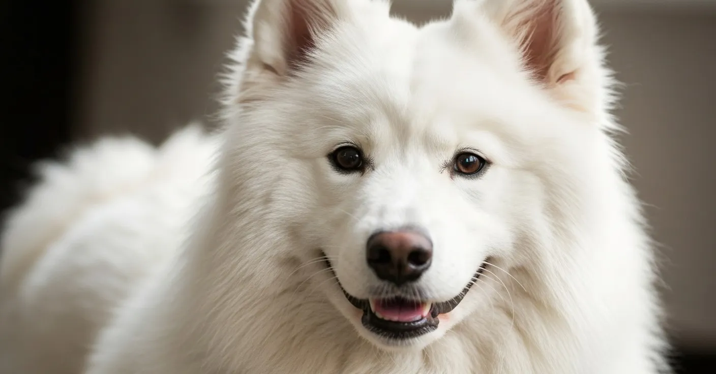
Samoyed
The Samoyed is an outlier on this list in one sense: they’re not brachycephalic at all. The skull is long, the face is open, and the nasolacrimal anatomy is normal. So why are they here?
Two reasons. First, the Samoyed coat is among the most dramatically white of any breed — pure, bright white with very little yellow or cream — which means any staining shows with maximum contrast. Second, Samoyeds have moderately prominent eyes for a spitz-type breed, and they’re often kept in outdoor environments where allergen exposure is high. Grass pollen, tree pollen, environmental molds — all of these can trigger low-grade allergic inflammation that elevates tear production and porphyrin output. The staining in Samoyeds tends to be more responsive to environmental allergen management than in brachycephalic breeds, precisely because the underlying driver is often allergic rather than structural.
What makes one dog in the same breed stain worse than another
This is one of the most common questions owners have after learning their breed is predisposed. Two dogs from the same litter, same diet, same household — and one stains heavily while the other barely shows a mark. What’s going on?
Several variables interact.
Genetics. Tear duct anatomy varies within breeds, not just between them. One Maltese may have slightly better nasolacrimal drainage than another from the same litter. Baseline porphyrin levels in tears are also heritable, at least partially.
Water source. High iron content in tap water provides additional substrate for porphyrin synthesis. In households where one dog drinks exclusively from a shared bowl filled from the tap and another has filtered water — or where the mineral content of local water changes over time — staining can diverge between otherwise identical dogs.
Diet composition. Iron-rich diets, particularly those heavy in organ meat, may support higher porphyrin production in some individuals. The response varies by dog.
Allergen exposure. A dog with a grass allergy living in a house with a large lawn is going to have chronically higher baseline inflammation — and likely higher tear volumes — than a genetically identical dog in an apartment.
Stress. Physiological stress increases cortisol and triggers inflammatory cascades. Dogs in high-stimulus environments, recently relocated dogs, and dogs with separation anxiety sometimes show dramatically increased staining that correlates with their stress load rather than their diet or water.
Managing expectations by breed
Honest breed-by-breed expectations matter because they shape how owners approach the problem.
Shih Tzu owners should expect tear staining to be a permanent feature requiring consistent management — daily face cleaning, weekly coat trimming around the eyes, and a long-term approach to internal porphyrin support. The structural anatomy doesn’t change. What changes is how well the visible staining is managed.
Maltese owners often find that consistent supplementation combined with diligent grooming produces meaningful results, particularly if dietary and water adjustments are made. The Maltese’s staining is less anatomy-driven than the hard brachycephalics, which means there’s more room for intervention to make a visible difference.
Poodle owners with affected individuals frequently see good responses to dietary changes and supplementation specifically because the epiphora in Poodles is often production-driven rather than drainage-impaired. Reducing porphyrin load in the tear film can show visible results in new coat growth within six to eight weeks.
Samoyed and Westie owners may find that addressing allergen exposure — antihistamines, allergen management, air filtration, foot washing after outdoor time — produces improvements that pure tear stain supplementation alone wouldn’t achieve, precisely because allergens are often the primary driver.
For all breeds, the framework that consistently produces the best outcomes combines external care (keeping the periocular area dry and trimmed) with internal support targeting the three main staining pathways: porphyrin production, oxidative discoloration, and microbial amplification.
The management framework
Daily external care is the foundation. Keeping the periocular fur trimmed short reduces the surface area available for porphyrin deposition and oxidation. Cleaning with a gentle, dog-safe solution at the eye margin daily removes fresh deposits before they oxidize and set. Drying the area thoroughly after cleaning matters — the warm moist environment that encourages Malassezia growth doesn’t get a chance to establish if the fur is kept dry.
Internal support complements this by addressing the upstream cause rather than just the downstream residue. Petterm Tear Stain Powder provides lactoferrin (which binds free iron, reducing porphyrin substrate availability, and inhibits the microbial species that intensify staining), lutein (an antioxidant that may help reduce oxidative discoloration in deposited porphyrins), and probiotics (which support healthy gut microbiome balance and help maintain lower systemic inflammation). It’s formulated as a daily powder that mixes into food, making it practical for long-term consistent use.
Consistency is the operative word. A single week of wiping and supplementing won’t produce visible changes. Eight to twelve weeks of consistent routine — external care daily, supplementation daily — is the minimum timeframe for seeing new unstained coat growth emerge and the old stained hair grow out. For the science behind what porphyrin is and why this timeline works the way it does, see our article on what porphyrin is and how it causes staining. For the complete picture of causes and solutions, visit our full guide on tear stain causes and solutions.
A note on cats
If you’re reading this as a multi-pet household that includes cats, the same visibility logic applies. Persian and Himalayan cats — both heavily brachycephalic — develop tear staining for precisely the same anatomical reasons as Shih Tzus and Bichons: compressed nasolacrimal drainage combined with prominent eyes and pale fur. The porphyrin chemistry is identical. The management principles are similar. If you’re managing staining in a white-coated dog and want to understand the same issue in your Persian or Himalayan, the tear stain causes and solutions guide covers the feline picture as well.
Frequently asked questions
My Maltese stained heavily as a puppy but improved at about a year old. Is that normal? Yes. Puppies often produce higher porphyrin levels and have less mature nasolacrimal drainage than adult dogs. Many white-coated breeds show peak staining in the first year and then improvement as the adult drainage system matures. This isn’t universal — some dogs maintain heavy staining into adulthood — but it’s a common pattern.
Is there a genetic test for tear staining predisposition? Not currently for staining specifically. Some genetic health panels test for conditions that can cause epiphora secondarily, but there’s no direct genetic marker for porphyrin excretion levels in commercial veterinary testing.
Does the color of a Poodle’s coat matter? My apricot Poodle stains more than my friend’s black one. The staining is equally present on both dogs biochemically. The apricot and light cream coats make porphyrin discoloration visible; the dark coat hides it. Your apricot Poodle doesn’t have more staining in an absolute sense — you can just see it.
I’ve heard that certain eye discharge types indicate infection. What should I look for? Clear, watery discharge is typical of normal tearing and porphyrin-related staining. Thick, opaque, yellow, or green discharge suggests active infection and warrants prompt veterinary attention. Discharge that changes suddenly in volume or character — in a dog with previously stable staining — is also worth a vet visit.
Can a Shih Tzu’s tear staining be permanently solved? Not given current veterinary options without surgical intervention to improve drainage, and even surgical dacryocystorhinostomy doesn’t guarantee complete resolution. The goal for most Shih Tzu owners is effective ongoing management, not a one-time fix.
My Westie’s staining gets much worse every spring. Why? Westies have a high rate of atopic dermatitis, and grass and tree pollen are common triggers. Spring allergen exposure drives inflammation, which in turn affects tear production and porphyrin levels. If the pattern is very consistent seasonally, it’s worth discussing environmental allergen management with your vet.
At what age should I start stain management in a new puppy? External grooming — keeping the face clean and dry — can start from the first weeks. For supplements like Petterm Tear Stain Powder, check the product guidance and consult your veterinarian, particularly for puppies under 12 weeks old.
Is staining in a Samoyed ever a sign of something more serious? Samoyeds don’t typically have structural drainage impairment, so sudden-onset or unilateral staining in a Samoyed is worth investigating more promptly than in a brachycephalic breed where structural causes are expected. The same rule applies as for any breed: bilateral stable staining in a predisposed dog is usually benign; sudden, unilateral, or changing staining warrants evaluation.
When to contact your veterinarian
Watch at home: Bilateral, mild, stable staining in a known stain-prone breed — especially brachycephalic breeds or any of the white-coated breeds described above. This is the normal expected presentation. Support with daily grooming, environmental and dietary adjustments, and consistent supplementation.
Call your vet within 24–48 hours: Sudden onset of significant staining in an adult dog with no prior history of it. Staining in one eye only (unilateral) — asymmetric staining can indicate a localized cause on that side, such as nasolacrimal obstruction, a foreign body, or early eyelid abnormality. Any change in discharge character: increased volume, thicker texture, or color shift from clear toward yellow or gray.
Seek same-day care: Green or yellow discharge, which strongly suggests active bacterial infection. Any change in corneal clarity — cloudiness, haziness, or a blue-white film over the eye. Squinting, light sensitivity, or your dog holding the affected eye closed. Visible swelling of the eyelid or tissue around the eye. Your dog persistently pawing at or rubbing their face against furniture — this can rapidly worsen corneal abrasion.
References
- Gelatt, K.N., Gilger, B.C., & Kern, T.J. (2013). Veterinary Ophthalmology. 5th ed. Wiley-Blackwell.
- Maggs, D.J., Miller, P.E., & Ofri, R. (2013). Slatter’s Fundamentals of Veterinary Ophthalmology. 5th ed. Elsevier.
- Petersen-Jones, S. & Crispin, S. (2002). BSAVA Manual of Small Animal Ophthalmology. 2nd ed. BSAVA.
- Bedford, P.G.C. (1999). Diseases and surgery of the canine nasolacrimal apparatus. In: Gelatt KN, ed. Veterinary Ophthalmology. 3rd ed. Lippincott Williams & Wilkins.
- Stades, F.C., et al. (2007). Ophthalmology for the Veterinary Practitioner. 2nd ed. Schlütersche.
This article is for educational purposes and is not veterinary medical advice. Petterm products are not intended to diagnose, treat, cure, or prevent any disease. Results may vary. Always consult your veterinarian before introducing a new supplement, especially if your pet has an existing eye condition, takes medication, is pregnant, or is under 12 weeks old.