


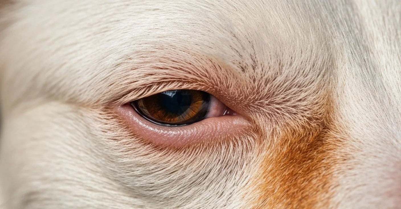
A client brought her white Maltese into the clinic absolutely convinced the dog had an eye infection. The fur beneath both eyes had turned a deep rust-brown — almost mahogany — and the owner had been applying warm compresses every morning trying to clear it up. Her groomer had suggested it might be bacterial. A neighbor told her to try boric acid. She’d been worrying about it for six weeks.
The examination took about three minutes. No discharge. No redness of the sclera. No squinting. The eyes themselves were perfectly healthy. What she was looking at wasn’t an infection at all. It was porphyrin — a naturally occurring molecule that dogs excrete through their tears — and it had been sitting on that white coat long enough to oxidize into something that genuinely looks alarming if you don’t know what you’re seeing.
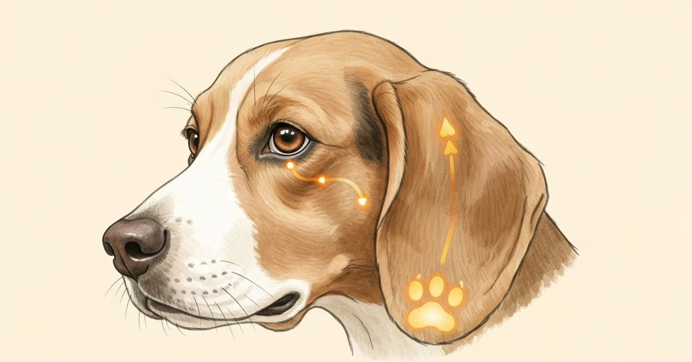
Understanding porphyrin doesn’t just solve a cosmetic problem. It changes how you approach the whole question of tear staining — what’s worth treating, how to treat it, and what realistic results look like over time.
What porphyrin actually is
Porphyrin isn’t a bacterium, a fungus, or a toxin. It’s a molecule — specifically, a tetrapyrrole ring structure that binds iron at its center. The most familiar porphyrin in biology is heme, the iron-containing component of hemoglobin that allows red blood cells to carry oxygen through the body.
As red blood cells age and are broken down — a normal, continuous process in every mammal — the heme is dismantled. The iron gets recycled and the tetrapyrrole ring is metabolized into a family of compounds collectively called porphyrins. They’re a byproduct of red blood cell turnover, not a sign that something has gone wrong (Gelatt et al., 2013).
The body has several routes for eliminating these breakdown products. In dogs, a meaningful portion exits through the tear film, through saliva, and through urine. This is why you can sometimes see rust-colored staining at the corners of a dog’s mouth, on the fur between the toes (where dogs lick), and even on the belly. It’s the same molecule taking the same biological exit — just landing on a different patch of fur.
How dogs excrete porphyrin
The lacrimal glands produce tear film continuously. Tear film isn’t just salt water — it’s a layered mixture of water, lipids, and proteins whose job is to keep the corneal surface moist, remove debris, and deliver oxygen to the eye. Porphyrins travel dissolved in the aqueous layer of this film.
Under normal circumstances, most of that tear film drains into the nasolacrimal duct: a narrow channel that runs from the inner corner of each eye down through the bone of the face and empties near the nostril. When drainage works well, the tears move efficiently away from the ocular surface and relatively little sits on the fur for long.
When drainage is impaired — or when tear production is simply higher than the drainage system can handle — tears overflow onto the face. That overflow carries porphyrins with it, and those porphyrins land on the periocular hair where they’re exposed to light, heat, and time. The result is staining (Maggs et al., 2013).
Saliva tells a parallel story. Dogs lick their paws. They groom their faces. Porphyrin-rich saliva deposits the same compound onto fur at every point of contact, which is why the rusty staining between the toes is often called “salivary staining” even though the underlying molecule is the same one responsible for tear stains. One molecule, multiple exit points.
Why the stains turn rust-red
Fresh porphyrin isn’t particularly dramatic in color. Porphyrins have some natural pigmentation, but in solution — dissolved in tears — they don’t look like much. What produces the deep, saturated rust-brown color that pet owners find so striking is oxidation.
When porphyrins are exposed to oxygen and UV light, they undergo photodegradation and oxidative reactions that produce increasingly darkened pigments. This is a well-documented property of porphyrin chemistry and explains a few things that owners often find confusing.
First, it explains why the staining is always worse on white or pale coats. The chemistry is identical on a dark-coated dog — the porphyrins are there, the oxidation is happening — but dark pigmented fur simply masks the discoloration. You can’t see rust-brown on chocolate brown (Petersen-Jones & Crispin, 2002).
Second, it explains why stains intensify over time even when tear production hasn’t increased. More UV exposure, more oxidation, darker color. A dog who spends hours outdoors in sunlight will often stain more visibly than an indoor dog with identical tear chemistry.
Third — and this matters for setting expectations — it explains why existing stains don’t fade when you address the underlying cause. You’re stopping new porphyrin from depositing on new fur growth. The already-stained hair has already been oxidized. It will grow out and be trimmed away. That’s the realistic timeline, and we’ll come back to it.
What drives higher porphyrin production
Not every dog produces the same volume of porphyrin in their tears. Several factors push production higher, and understanding them opens the door to meaningful intervention.
Iron-rich diet and water. Because porphyrins are iron-binding molecules derived from heme breakdown, higher iron availability means more substrate for porphyrin synthesis. Diets with very high meat content, particularly organ-heavy raw diets, may elevate porphyrin levels in some dogs. Water source is a frequently discussed variable — high-mineral water with elevated iron content may contribute to increased porphyrin excretion, though rigorous controlled trials in companion dogs on this specific question are limited.
Red blood cell turnover rate. Any condition that accelerates the breakdown of red blood cells will increase porphyrin output. Systemic infection, chronic inflammation, immune-mediated hemolysis, and even prolonged physical stress can push RBC turnover higher than baseline, meaning more heme is being processed and more porphyrin is entering the excretory pathways (Gelatt et al., 2013).
Stress. Physiological stress elevates cortisol and triggers systemic inflammatory cascades that can secondarily affect RBC lifespan. Shelter dogs, recently rehomed dogs, and dogs in high-stimulus environments often show increased tear staining during periods of adjustment — owners sometimes misinterpret this as a dietary problem when the driver is psychological.
Age and hormonal status. Puppies, especially those under a year old, often stain more heavily than adult dogs. Hormonal shifts around the first heat cycle in females can also temporarily increase staining.
The difference between staining and epiphora
These two terms often get conflated, and they shouldn’t be. Staining is a visible residue. Epiphora is a condition — specifically, excessive tear overflow onto the face, either because the eye is producing too many tears or because drainage is impaired.
Staining can occur without clinical epiphora: a dog with normal tear production and intact drainage but elevated porphyrin concentration in those tears will still stain. Epiphora can occur without dramatic staining: a dog with very pale, low-porphyrin tears and a dark coat may overflow visibly but show no discoloration.
In practice, the two coexist frequently. Epiphora deposits more porphyrin-containing fluid onto the periocular fur than normal drainage would, and that additional exposure means more substrate for oxidation. Identifying whether epiphora is present matters clinically because it may signal a structural issue — nasolacrimal obstruction, eyelid abnormalities, corneal irritation — that warrants veterinary evaluation rather than just supplementation (Maggs et al., 2013).
Why breed anatomy matters
In brachycephalic dogs — the breeds with compressed facial anatomy like Bulldogs, Shih Tzus, Pugs, and Pekingese — the nasolacrimal duct often runs a distorted or partially obstructed course through the shortened skull. The duct was designed for a longer face. When that face gets compressed through selective breeding, the duct gets kinked, narrowed, or angulated in ways that impair flow.
The result is structural epiphora. Tears that should drain internally instead spill over the lid margin and run down the face. This isn’t something that dietary changes or supplements will structurally correct — but it does mean the management of porphyrin concentration in those tears becomes more important, because more tear volume is reaching the fur.
Prominent, slightly exophthalmic (forward-set) eyes common in brachycephalic breeds also expose more of the ocular surface to air, which increases evaporation and may stimulate reflex tearing as compensation. More tearing, more porphyrin delivery to the periocular region, more staining potential.
Non-brachycephalic breeds with normal skull anatomy can also stain heavily, but their staining is more likely driven by porphyrin concentration itself, dietary factors, or early-stage nasolacrimal obstruction rather than structural overflow.
Microbial amplification
Porphyrin alone accounts for a significant portion of tear stain discoloration. But it doesn’t account for all of it — and in dogs with severe, very dark staining, secondary microbial activity often plays an amplifying role.
The periocular fur in a dog that tears chronically is warm and consistently moist. That environment is highly hospitable to Malassezia species — the yeast genus associated with skin and ear problems in dogs — and to various bacterial species. These organisms colonize the hair shaft and the skin beneath it, and their metabolic byproducts include additional pigmented compounds that stack on top of porphyrin-driven discoloration.
The net result can be staining that’s considerably darker and more saturated than porphyrin oxidation alone would produce. This explains why dogs with similar tear volumes can show dramatically different stain severity: one dog’s periocular environment may have heavy Malassezia colonization while another’s stays relatively clear (Petersen-Jones & Crispin, 2002).
External grooming matters here. Keeping the periocular area dry and clean reduces the substrate available for microbial growth. But addressing the microbial component externally while ignoring the upstream porphyrin volume is fighting on one front while ignoring the other.
Internal approaches to porphyrin load
This is where the evidence base for supplementation becomes relevant. Several compounds have plausible mechanisms for reducing either porphyrin-driven staining specifically or the oxidative discoloration it produces.
Lactoferrin is an iron-binding glycoprotein found naturally in milk, tears, and saliva. It has a high affinity for iron, and by sequestering free iron in the gut and in secretions, it may reduce the availability of iron for porphyrin synthesis. Less free iron circulating means potentially less substrate driving porphyrin production. Lactoferrin also has well-documented antimicrobial properties — it’s been shown to inhibit bacterial and fungal growth through iron deprivation and direct membrane disruption (Farnaud & Evans, 2003; Giansanti et al., 2016). This dual mechanism — reducing porphyrin substrate and inhibiting the Malassezia and bacterial species that amplify staining — makes it one of the more mechanistically coherent ingredients in the tear stain supplement space.
Lutein is a carotenoid antioxidant. Since porphyrin discoloration is partly driven by oxidation, antioxidant compounds that reduce oxidative activity in tear secretions may help maintain a lighter color in deposited porphyrins. Lutein is well established in the human ophthalmological literature as a compound that concentrates in ocular tissue, and its use in veterinary eye health supplements follows from that evidence base.
Probiotics work more indirectly. Gut microbiome composition influences systemic inflammation, and systemic inflammation affects RBC turnover rates and, by extension, porphyrin production. Probiotic supplementation that helps maintain healthy gut bacterial populations may contribute to lower background inflammation and a more stable porphyrin output.
Petterm Tear Stain Powder combines lactoferrin, lutein, and probiotics in a daily powder formulation designed to address these three pathways together. For a broader look at what causes tear staining and how to address it from multiple angles, see our full guide on tear stain causes and solutions.
What to expect realistically
If you begin addressing porphyrin load through diet adjustment, water source changes, and supplementation, the first thing to know is that the existing stained fur isn’t going anywhere on its own. Oxidized porphyrin is deposited in the hair shaft. It doesn’t un-oxidize. No supplement fades existing staining.
What changes with effective intervention is new growth. The fur growing in from the follicle — the hair that wasn’t there when you started — won’t carry the same staining if porphyrin levels have been meaningfully reduced. Over four to eight weeks, depending on coat growth rate, you’ll typically start to see a lighter band of new growth near the skin. The old stained hair grows out, gets trimmed, and is replaced by unstained growth.
This timeline is frequently misunderstood, and it’s the main reason owners abandon supplements too early. Four weeks in, the staining still looks the same — because the old stained fur is still there. Eight to twelve weeks in, with the coat growing out, the difference becomes visible.
For more information on which white-coated breeds are most affected and what management looks like by breed, see which white-coated breeds are most affected.
Frequently asked questions
Is porphyrin dangerous to my dog? No. Porphyrin is a normal metabolic byproduct of red blood cell breakdown, and its presence in tears is entirely physiological. The staining it produces is cosmetic — it doesn’t harm the eye or the skin beneath the fur in healthy dogs.
Why does my dog stain worse after being outdoors? UV light accelerates the oxidation of porphyrins. More sun exposure means faster and darker oxidation of the porphyrins already deposited on the fur. The chemistry isn’t different; the reaction is just faster.
Can I remove the staining with topical products? Some topical whitening solutions can lighten existing stains modestly. Most work by bleaching rather than removing porphyrin. They don’t address the source. For a lasting change, reducing porphyrin deposition is more effective than bleaching what’s already stained.
Does the water my dog drinks really matter? Water mineral content — particularly iron — is frequently cited as a contributing factor. Some owners report improvement after switching to filtered or distilled water. The evidence in companion animals is largely observational rather than controlled, but it’s a low-risk variable worth adjusting if staining is severe.
My dog’s saliva is staining their paws too. Is this the same thing? Yes. Porphyrins are excreted through saliva as well as tears. The reddish-brown staining between the toes in dogs who lick their paws is salivary porphyrin staining, and the same interventions that reduce tear staining may also reduce salivary staining over time.
How long before I see results from supplementation? Four to eight weeks is a realistic range for seeing visible new unstained growth, depending on how fast your dog’s coat grows. Existing stained fur doesn’t fade — the improvement is in new growth replacing old.
Is porphyrin staining worse in some seasons? Some owners notice seasonal variation, likely related to UV exposure and allergen-driven inflammation. Spring and summer tend to correlate with more pronounced staining in allergy-prone dogs.
Can porphyrin staining indicate a health problem? In most cases it’s normal variation. Sudden increases in staining, especially in an adult dog with no prior staining history, are worth investigating because they could signal increased RBC turnover from an underlying condition. Unilateral staining (one eye only) warrants a vet visit to rule out localized causes.
When to contact your veterinarian
Watch at home: Bilateral, mild, chronic staining that has been stable in a known stain-prone breed — especially a brachycephalic or white-coated dog. Support with consistent grooming, water quality management, and supplementation. This is the normal picture for breeds like Maltese, Bichon Frise, and Shih Tzu.
Call your vet within 24–48 hours: Sudden onset of staining in an adult dog with no prior history. Staining in only one eye (unilateral), which may indicate a localized issue such as nasolacrimal obstruction, eyelid abnormality, or foreign body on that side. Discharge that has changed in character — more volume, different color, or thicker texture than before.
Seek same-day care: Green or yellow discharge from the eye, which suggests active bacterial infection. Any cloudiness, haziness, or change in the appearance of the cornea or lens. Persistent squinting or sensitivity to light. Visible swelling around the eye or eyelids. Your dog pawing at or rubbing their face repeatedly — this can worsen corneal damage quickly.
References
- Gelatt, K.N., Gilger, B.C., & Kern, T.J. (2013). Veterinary Ophthalmology. 5th ed. Wiley-Blackwell.
- Maggs, D.J., Miller, P.E., & Ofri, R. (2013). Slatter’s Fundamentals of Veterinary Ophthalmology. 5th ed. Elsevier.
- Farnaud, S. & Evans, R.W. (2003). Lactoferrin: a multifunctional protein with antimicrobial properties. Molecular Immunology, 40(7), 395–405.
- Petersen-Jones, S. & Crispin, S. (2002). BSAVA Manual of Small Animal Ophthalmology. 2nd ed. BSAVA.
- Giansanti, F., et al. (2016). Lactoferrin from milk: nutraceutical and pharmacological properties. Pharmaceuticals, 9(4), 61.
This article is for educational purposes and is not veterinary medical advice. Petterm products are not intended to diagnose, treat, cure, or prevent any disease. Results may vary. Always consult your veterinarian before introducing a new supplement, especially if your pet has an existing eye condition, takes medication, is pregnant, or is under 12 weeks old.